
Choledochal cyst
in children

A choledochal cyst is a swelling or widening of the bile ducts. They are rare and are treated with surgery.
Also called: Choledochal anomaly, choledochal malformation
The information on this page is for:
- Parents of children with choledochal cyst or supected choledochal cyst.
- Young people with choledochal cyst.
- Family, friends, carers, and healthcare professionals.
Find out more about choledochal cysts in adults
How to say it: col-eh-DOKE-al / sist
On this page:
Quick Summary
- Choledochal cysts are very rare.
- They are swellings in the bile ducts that start before you are born.
- The condition is usually diagnosed in children.
- Choledochal cysts are treated with surgery.
Find out more about choledochal cysts in children on this page.
Useful words
Bile A liquid produced in the liver that helps digest fat
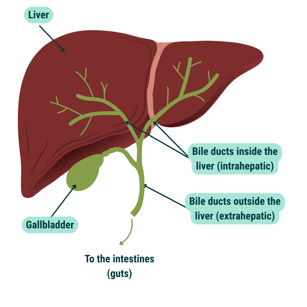
Bile ducts The tubes that carry bile from the liver to the gallbladder and intestines
Biliary tree Another name for bile ducts
Intrahepatic bile ducts The bile ducts that are inside the liver
Extrahepatic bile duct The bile duct that is outside the liver
Common bile duct Another name for the bile duct that is outside the liver
Gallbladder A sac where bile is stored before going to the intestines
What is a choledochal cyst?
The term choledochal cyst can be broken down into the following:
- chole ~ relating to bile
- dochal ~ containing or receiving
- cyst ~ fluid collection
A choledochal cyst is a swelling / widening or dilatation of the bile ducts. The condition usually affects the part of the bile ducts outside the liver (the common bile duct and the hepatic ducts) but sometimes it also affects those inside the liver (the intrahepatic ducts).
We don’t know the exact cause of a choledochal cyst but there are a few theories. One explanation is that it happens due to the bile duct not forming properly when the baby is developing in the womb in early pregnancy. This concept is supported by the fact that in some cases, the cyst is first seen during a routine ante-natal ultrasound scan of the baby in early pregnancy.
Another theory is that the junction of the bile duct with the pancreatic duct is not normal and forms an abnormally long ‘common channel’. Currently there are arguments in support of both of these.
Bile and bile ducts
Our liver produces a secretion known as “bile” which contains useful products. Bile has many functions which help in food absorption from the gut. Bile also gets rid of waste products that are not needed by the body.
Bile ducts are tubes which carry bile from the liver to the gall bladder and then to the gut when required.
Is the condition rare?
Yes, it is rare. It affects about 1 in 50,000 children in the West. Choledochal cysts are three times as common in girls as boys and rarely run in families.
What are the different types of choledochal cyst?
There are several types of choledochal cyst. Their positions and sizes may differ.
Type 1
Type I cysts are the most common. The ducts inside the liver look normal but the common bile duct may be swollen up like a small balloon or a fat cigar.
In these types of choledochal cyst, the lower end of the common bile duct often joins up with the pancreatic duct. This is not normal and forms an abnormally long ‘common channel’. This causes the pancreatic juice and the bile to mix together very easily before they enter the duodenum. This early mixing can
result in weakening of the bile duct wall leading to ballooning.
In addition it can lead to complications such as pancreatitis (inflammation of the pancreas) or cholangitis (inflammation of the biliary system).
Other types
Other types of choledochal cysts are less common:
- Type 2
- Type 3
- Type 4a
- Type 5
Apart from these types of choledochal cyst there is another known as the “forme fruste” type of choledochal anomaly. Although this involves an abnormality of the junction between the bile duct and pancreatic duct there is no widening (dilation) of the ducts. This kind of abnormality can also give rise to serious
complications.
What are the signs and symptoms of a choledochal cyst?
Some of these cysts are found before birth on the antenatal scan. Sometimes, a choledochal cyst is found when a child is having an abdominal ultrasound scan for an unconnected reason like pain in the abdomen. In some cases the child may have symptoms such as jaundice, a mass in the abdomen, pancreatitis or cholangitis.
Most choledochal cysts are detected in childhood. A child with a choledochal cyst may have some or possibly all of the following symptoms:
- Jaundice – This is due to blockage of bile drainage. This might be prolonged jaundice (lasting more than two weeks) in a newborn baby or intermittent jaundice (jaundice which comes and goes) in an older infant or child. Usually, the jaundice is associated with pale coloured stools and dark urine.
- Intermittent abdominal pain (abdominal pain which comes and goes) which can be severe
- Cholangitis (an infection of the bile duct)
- Peritonitis (a severe inflammation within the abdomen) if the cyst bursts or leaks
- A lump or swelling in the belly
- Pancreatitis
- An inflammation (irritation, swelling and damage) in the pancreas
How is a diagnosis made?
The diagnosis is generally made using a combination of the following:
- History
- A physical exam of the child
- Tests including blood tests and an ultrasound scan
A choledochal cyst is most often suspected if an abdominal ultrasound scan shows a dilated bile duct in a baby or child with jaundice or if the child has abdominal pain. Once suspected some other tests are required to get more information. For example, is there any blockage in the duct by a stone or is there an abnormal junction of the bile duct with pancreatic duct (a long common channel)?
These extra tests are carried out to get more information about the type of choledochal cyst present which helps the surgeon to get a ‘road map’ to plan the operation.
These tests may include:
Ultrasound scan
A more detailed ultrasound scan to look not only at the bile ducts but also to look at the blood vessels going to the liver which run alongside the bile ducts.
MRCP (Magnetic Resonance Cholangio-Pancreatography)
This type of detailed scan is carried out in a large machine. It does not require X-rays but the patient does need to lie flat and still and so babies and small children often require a general anaesthetic. The bile ducts and the pancreatic duct are shown better on an MRI scan.
ERCP (Endoscopic Retrograde Cholangio-Pancreatography)
This is a specialised procedure which is not often needed. It is done under a general anaesthetic.
In this procedure a flexible tube with a small camera on the end (an endoscope) is passed through the mouth down into the intestine. A tiny plastic tube is then passed through this endoscope into the opening of the bile duct and dye is injected to outline the choledochal cyst and the pancreatic duct. This helps to give information about the junction of bile duct with pancreatic duct (long common channel) and if there is any blockage in the bile duct due to stones etc.
This investigation can sometimes stir up inflammation in the pancreas (pancreatitis) causing temporary abdominal pain and vomiting afterwards. It is rarely done as it is an invasive investigation.
HIDA (liver scan)
Rarely a HIDA (liver scan) becomes necessary, if there is only a single cyst in the liver and the MRCP cannot confirm it to be a choledochal cyst/anomaly.
The exact nature of the choledochal cyst sometimes only becomes clear at the time of surgery when dye is injected into the bile duct system (an operative cholangiogram).
What is the treatment for a choledochal cyst?
The treatment of choledochal cysts is surgery. This should be done in a centre which has experience of looking after children who have choledochal cysts and surgeons experienced with this kind of operation.
The operation can be performed as key hole surgery (using a small opening through the skin) or as an open procedure (a larger opening in the skin). Both techniques achieve good results.
In the common variety, the cyst is completely removed. This means removing most of the bile ducts (tubes carrying bile) outside the liver together with the gallbladder. The hepatic ducts (tubes from the liver) coming out of the liver are then attached to a loop of the child’s bowel so that the bile can drain into the bowel. This loop of bowel is a specially created loop. Removing the gallbladder has no important long-term effects that we know of. A liver biopsy may also be taken at the time of surgery to see if the liver is healthy or damaged.
In babies with a cyst discovered by ultrasound scan but who have no symptoms, there is debate about the best time to perform surgery. Children’s surgeons who specialise in liver and bile duct surgery often recommend early surgery, generally at about six months of age.
Are there any complications of surgery?
There are risks as with any other surgery but these are not common. For example, there is a small risk of bleeding, and infection. There is also a risk of leakage and narrowing where the bile ducts are joined to the bowel.
The operation is generally very successful. Most of the time, children recover rapidly and are able to go home within one week after surgery. They are then seen in the outpatient clinic from time to time when a blood test (to check liver function) and an ultrasound scan may be performed.
What about the long term outlook after surgery?
The majority (90% or more) of children remain well and grow up to be healthy adults. A small number (10% or less) may get problems such as cholangitis. Cholangitis is an infection of the bile ducts within the liver. If it occurs it generally happens in the first few months after surgery. If a child becomes unwell after surgery e.g. has a temperature, it is important to seek medical advice.
Some children require further surgery, for example, because the joint between the bile ducts and the bowel has narrowed down. In a very small number of patients, there is a small, long term risk of other complications including cancer of the bile ducts in adult life. This is extremely rare if the cyst has been removed properly (not leaving behind a portion of cyst or delaying the surgery for long time).
Rarely, the liver is very scarred (cirrhosis) when a choledochal cyst is first diagnosed. In such cases, medical monitoring is necessary after the cyst has been removed because further problems can develop and these need to be recognised early.
What happens if a choledochal cyst is left untreated?
If a choledochal cyst is left untreated, it may lead to major complications. These can include:
- Liver damage (fibrosis and cirrhosis)
- Infection (cholangitis)
- Bile duct damage
- The cyst may burst or leak
- Problems with food absorption and growth
- Bleeding
- Stone formation (gallstones) in the cyst
- Pancreatitis (inflammation in the pancreas) and damage to the pancreas and its duct
- Risk of cancer developing within the bile tubes or in the gallbladder
Your specialist liver team will discuss the risks to your child if the cyst is left untreated.
Support
How Liver UK can help
A diagnosis of liver disease can be worrying, and you may have a lot of questions.
We're here for you and for your family and friends. Whether you have questions or just need someone to listen, we can help.

Your feedback
This content was last reviewed: December 2020
Our expert reviewers:
We would like to thank everyone who helped with creating and reviewing this page. This information has been produced with input from the three specialist paediatric liver centres in the UK. And with parents and families.
Find out how we make our patient information.
Everyone’s experience of liver disease will be different. Always talk to your specialist medical team for personal advice.
More from Liver UK

Check your risk
Find out more

Support us
Find out more
Keep up to date
Find out more